|
FEATURE
From Photo Digitization to OER
by Bryan Hull, Carmin I. Smoot, and Julia Curtis
| The scope of the project quickly expanded beyond a simple flash card system to a platform that could allow users to explore the image set via diagnosis and clinical characteristics. |
 |
This is the story of how a photo digitization project is transforming into a library-developed OER for doctors, students, and researchers.
Background of the UtahDERM project
In fall 2017, Dr. Garrett Coman, a dermatology resident at the University of Utah, asked the Spencer S. Eccles Health Sciences Library to digitize a collection of nearly 15,000 35-millimeter Kodachrome slides belonging to Dr. Leonard Swinyer, an adjunct faculty member for the University of Utah’s department of dermatology. After more than 4 decades working in dermatology, Swinyer had amassed an extensive clinical image collection of patients’ skin conditions. Taken with patient permission, this high-quality clinical image collection was significant in terms of its size, breadth, and educational value, with coverage of both pediatric and adult patients, as well as a representation of skin colors to reflect the diversity of the patient population.
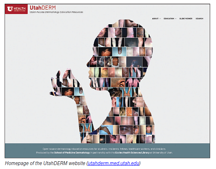
When Coman approached the library with the slides, the initial aim was to digitize the images for preservation; later, they could be turned into digital flash cards to further the education of medical students and residents. The scope of the project quickly expanded beyond a simple flash card system to a platform that could allow users to explore the image set via diagnosis and clinical characteristics. Thus, the seeds were planted for the UtahDERM (Dermatology Education Resources & Modules) project.
Digitizing the Kodachrome Slides
Utilizing a CyberView X5-MS automated batch scanner, each Kodachrome slide was scanned at 400 DPI. The resolution was chosen for the speed with which the scans could be completed and for its suitability for web transmission and viewing. While a higher resolution is ideal for true digital preservation, it wasn’t feasible with the available resources and the desire to get the collection of images digitized quickly in order to begin work on the image viewer platform. In all, it took 988 hours of total effort to digitize the slides at the chosen resolution.
Metadata Structure and Gathering the Descriptions
To accurately describe the images, a standard was created for both diagnoses and clinical characteristics. For diagnoses, names were to match the dermatology textbook that the dermatology department uses for teaching and learning: Dermatology, Fourth Edition by Jean L. Bolognia, Julie V. Schaffer, and Lorenzo Cerroni (Elsevier 2018). This ensured that a single diagnosis name was used, reducing alternative names or aliases that are common in dermatology from being introduced into the dataset. Clinical characteristics, which refer to the visual information a dermatologist sees when observing a dermatologic condition, were also standardized in order to have a controlled vocabulary and standard with which to describe the images. The characteristics included the following:
- Location
- Configuration
- Color
- Primary lesion
- Secondary change
- Fitzpatrick I-VI
- Immunosuppression status
- Sex
The metadata gathering process was unique in that a team of 12 dermatology faculty members and residents was recruited to confirm the diagnosis and identify the clinical characteristics for each of the nearly 15,000 slides. The process for gathering metadata was as follows:
- A folder of images was assigned to a dermatology faculty member or resident.
- That person reviewed each image in the folder and filled out a Google Form with metadata.
- The process was repeated until the entire folder of images was described.
Each faculty member and resident received a small stipend for each image described, incentivizing their participation and compensating them for the significant time commitment necessary for such a large image set. The process to describe the images was monumental and took a year and a half to complete, with the first set of images being described in early 2019 and the final set being completed in July 2020.
Developing the Platform to Host and Share the Images
While the Eccles Library had access to readily available CMSs and digital collection solutions, none of them were equipped to handle the specifics of diagnosis and clinical characteristics that described the images. They were also unable to help us achieve the educational goals that were envisioned for the project. It became clear that a home-cooked solution needed to be developed by the library in order to make the vision of an educational tool and image explorer a reality.
A team of library personnel—including two librarians, a web programmer, and a web designer—began the development of the image viewer in fall 2018. The development followed standard user design prototyping practices, starting with low-fidelity prototypes and working toward a high-fidelity prototype that we could then use as the initial structure for the system. Feedback was sought along the way from our dermatology colleagues as we solidified design choices and explored features to be implemented.
There were four basic functionalities that were to be designed and implemented into the image viewer system:
- Browsing images by diagnosis
- Filtering the images by characteristics
- Browsing images via Dermatology textbook chapter
- Ability to hide diagnoses for flash cards
Each image had a dermatologic diagnosis that was ascribed through the metadata collection process. As such, the ability to search diagnoses was one of the primary functionalities of the image viewer; that way, users could explore entire image sets of the same diagnosis. We also wanted users to have the ability to search multiple diagnoses in order to compare image sets comprising discrete diagnoses.
To further refine image searching, we wanted to provide an option to filter the images by clinical characteristics. This included being able to search across the entire collection based on specific characteristics, such as location, but also to filter characteristics within specific diagnosis queries. For example, we wanted the ability to search all images that had conditions located on the hand and the ability to search a specific diagnosis, such as blue nevus, but to filter the search to only blue nevus that appears on the ears. This introduced complications into the system design, as the characteristics could act as discreet search terms but also as a filter to refine search returns of a diagnosis.
To increase the educational utility of the image viewer, an additional method to search the collection was introduced. This time, it was by chapter of the Dermatology textbook, in which a diagnosis is primarily referenced and discussed. This allows students and residents to browse images related to specific chapters within the text for study.
Lastly, the original idea for the system was to be able to flash-card the images. This proved to be complicated to implement, since a collection of images would first need to be queried before users could flash-card them. Thus, if the idea was to hide diagnoses in order to test your knowledge, you would not be able to search by diagnosis. Therefore, the only way to construct a query was via a clinical characteristic search in order to get an image set without revealing diagnoses. To work around this issue, a user can toggle between the two modes of the image viewer—search mode and flash card mode—to search for images, then flash-card the return.
The image viewer itself was built using many different technologies, including server-based PHP, several Java-Script libraries, HTML, CSS, and an IIIF image server to handle serving large images. The front end of the website was built on WordPress but is separate, as the image viewer runs as its own independent application. The back end of the image viewer contains systems for ingesting images, data management, and administrative tasks such as logging and auditing.
Medical Student Write-Ups
In conjunction with the development of the image viewer, a collection of articles covering 100 core dermatology diagnoses was developed as an additional feature of the UtahDERM project. These core diagnoses articles serve as a quick reference tool for medical students and general health practitioners to learn the essentials of common dermatological conditions. Each article is written by a rotating medical student in the department of dermatology and peer-reviewed by dermatology faculty members. Additionally, a linkback system was created in order to link diagnoses within the image viewer back to the relevant article and vice versa. This helped create additional learning opportunities to be included with the image viewer, as a user could reference basic information for common dermatologic conditions in conjunction with a large set of clinical images. It also presented an opportunity for medical students within the dermatology department to become involved in the project and gain experience with medical education, writing, and peer review.
Lessons Learned and Future of the Project
Now in our sixth year of the UtahDERM project, we have learned many valuable lessons for projects of this scope. The first challenge we encountered involved addressing the difficulty of the metadata gathering process, which necessitated bringing in subject matter experts. It is difficult to get highly specific metadata from a highly specialized group of people who have severe time constraints to describe an image set as large as the Swinyer collection, even with financial compensation as an incentive. It is also challenging to ensure quality control, as library personnel cannot judge the accuracy or completeness of the metadata and must rely on subject matter experts to peer review and perform quality control.
The second challenge involved tackling feature and scope creep in the development process, as the COVID-19 pandemic occurred in the middle of development and added another layer of complexity to project management. Connecting with our dermatology colleagues became more complicated, as meetings were canceled and more pressing issues rose to the forefront. For library personnel, this left extra time for development to focus on features and management structures that were not initially included in the original prototype and scope of the project.
While feature and scope creep presented challenges, they also offered opportunities to discover solutions that were ultimately beneficial to other projects, such as the inclusion of an IIIF image server that could be used elsewhere. Feature and scope creep also presented numerous areas of growth, as the platform itself has the flexibility to include other methods of dermatologic imaging, such as histopathology, dermoscopy, reflectance confocal microscopy in vivo imaging, and other modalities, as they arise in the future. The image viewer and website are currently in the alpha stage, meaning that the application and materials that are presently ingested are publicly accessible but not widely promoted or publicized yet.
Our future plans include completing the ingestion of the entire set of Kodachrome slides before a wider, more publicized release of the application. We are also hoping to recruit dermatology faculty members to provide more images in order to grow the collection, especially when it comes to adding more skin-of-color examples for each diagnosis. Lastly, we’d like to conduct more stringent user testing on the initial application so that we may further refine its user interface and streamline its functionality. Our hope is that with a wider release and more input from the dermatology community, we can grow the image set, find funding avenues to develop new facets to the image viewer, and have a widely used and helpful tool for dermatology education across the globe.
|